New Patients
Existing Patients
New Patients
Existing Patients
New Patients
Existing Patients
New Patients
Existing Patients

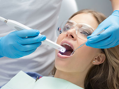
An intraoral camera is a compact, pen-sized imaging device that captures clear, full-color photos and video of the teeth, gums, and other soft tissues inside the mouth. Unlike a traditional dental mirror, this tool gives an enlarged, detailed view of small areas—cracks in enamel, the margins of restorations, early staining, and inflammation around the gumline. Images are displayed in real time on a chairside monitor so patients and clinicians can see the same view simultaneously.
The technology combines high-resolution optics with directed lighting to minimize glare and reveal subtle textures and color variations that are difficult to observe otherwise. Because it is minimally invasive and easy to maneuver, the intraoral camera can examine hard-to-see regions such as the back of the mouth, between teeth, and around dental work. The device’s small size makes it suitable for adult and pediatric patients alike and helps reduce discomfort during the exam.
Beyond simple observation, the camera can capture still images and short video clips that freeze a moment in time for closer study. Those visuals often make it easier to identify early-stage concerns that benefit from monitoring or conservative care. By providing a crisp, magnified view, the intraoral camera becomes a practical extension of the clinician’s eyes—enhancing precision without replacing clinical judgment or other diagnostic tools.
High-quality intraoral images strengthen diagnostic accuracy by revealing details that might be missed during a visual exam alone. Fine fractures, microleakage around fillings, and early decay tucked beneath enamel can all be easier to detect with magnified photos. When combined with a thorough clinical exam and radiographs where appropriate, these images contribute to a fuller understanding of a patient’s oral health and allow for more targeted treatment planning.
Because images can be captured from multiple angles, they help the dental team evaluate the condition of restorations and the fit of crowns, bridges, and orthodontic appliances. The ability to document a lesion or area of concern over time also supports conservative management strategies: clinicians can monitor stability or progression and make informed decisions about when intervention is truly necessary. This measured approach supports long-term oral health and avoids unnecessary procedures.
When treatment is indicated, intraoral photos assist in outlining options and sequencing care. Clear visuals make it easier to explain why a procedure is recommended and how it will address the observed problem. That clarity improves mutual understanding between clinician and patient, which in turn supports shared decision-making and increases the likelihood that care follows an evidence-based path rather than relying on guesswork.
One of the most valuable benefits of intraoral cameras is their ability to bridge the communication gap between clinician and patient. Instead of relying on technical descriptions, practitioners can show patients exactly what they see. This transparent approach helps patients grasp the nature and extent of an issue—such as a hairline crack or a small area of gum recession—so they can make better-informed choices about their care.
Seeing real images of their own mouth often reduces uncertainty and builds trust. Patients who understand the problem clearly are more likely to follow through with recommended home care and appointments. The visuals also serve as an educational tool; clinicians can point out proper brushing and flossing techniques, explain the implications of tissue changes, and demonstrate the effect of habits like bruxism or aggressive brushing in a way that words alone cannot convey.
For caregivers and families, especially those making health decisions for children or elderly relatives, intraoral images provide a useful reference. They function as a shared starting point during consultations, allowing the dental team to review findings, compare treatment alternatives, and outline realistic expectations. This collaborative communication supports better outcomes and a more comfortable patient experience.
Intraoral images are easily saved to a patient’s digital chart and organized as part of a permanent record. This documentation strengthens continuity of care by preserving a visual history that can be reviewed at future visits to assess changes or treatment response. Because images are dated and stored, clinicians can track healing, detect recurrence, and verify the long-term success of restorations or periodontal therapy.
When care requires consultation with a specialist or coordination with a dental laboratory, high-resolution photos streamline communication. Images can be shared securely with colleagues to illustrate a specific concern, ensuring remote collaborators see precisely what the referring clinician observed. Similarly, dental technicians use these visuals to match shades and shapes more accurately when fabricating crowns, veneers, or prosthetics, improving the fit and appearance of final restorations.
From a recordkeeping perspective, saved images support transparency and quality assurance. They help the dental team review clinical decisions and provide an objective reference should questions arise about treatment options or outcomes. In short, intraoral photos are an efficient way to connect clinical observation with the broader digital ecosystem that modern dental care relies on.
An intraoral camera exam is quick, noninvasive, and typically integrated into a routine checkup. During the exam, the clinician or hygienist will guide the small camera around the mouth while the patient sits comfortably in the chair. The procedure requires no special preparation, and most patients find it no more intrusive than routine dental instrumentation. The chairside monitor displays images in real time so the clinician can explain findings as they appear.
Because the camera uses visible light and optical lenses, there is no exposure to radiation during this part of the exam. The device is cleaned and sterilized between uses according to infection-control protocols, and single-use sleeves are often employed to further assure safety and comfort. Children and apprehensive patients benefit from the camera’s gentle approach—images are captured quickly and the clinician can stop at any time if the patient prefers.
After the exam, selected images may be saved to the chart for follow-up appointments and to document treatment needs or improvements. Clinicians typically use these visuals to outline next steps and answer questions, ensuring patients leave the appointment with a clear, visual understanding of their oral health. For many patients, seeing the condition of their own teeth and gums is a meaningful step toward proactive, informed care.
Intraoral cameras are a practical, patient-centered technology that enhances diagnosis, communication, and documentation without adding complexity to the visit. By providing enlarged, detailed images of teeth and soft tissues, these devices help clinicians spot issues earlier, explain conditions clearly to patients, and coordinate care with specialists and laboratories. The result is more precise care and a stronger partnership between clinician and patient.
If you would like to learn how intraoral imaging is used during routine exams or specialty appointments at RGV Smiles by Dr. Rocky L. Salinas, DDS, PA, our team is happy to explain the process and show examples during your visit. Contact us to request more information or to discuss what to expect at your next appointment.


An intraoral camera is a small, pen-sized imaging device that captures high-resolution still photos and short video of the teeth, gums and other soft tissues inside the mouth. It combines precision optics with directed lighting to reveal surface details such as hairline cracks, early decay and the margins of restorations that are difficult to see with the naked eye. Images appear on a chairside monitor in real time so clinicians and patients can review the same view together.
Because the device is minimally invasive and easy to maneuver, clinicians can examine hard-to-see areas like the back of the mouth and tight interproximal spaces with minimal discomfort. The camera’s design makes it suitable for adult and pediatric patients and helps reduce anxiety during exams. In clinical practice, intraoral imaging serves as an extension of the clinician’s visual exam, enhancing detail without replacing clinical judgment or other diagnostic tools.
High-quality intraoral images reveal subtle textures, color variations and microfractures that may be missed during a visual exam alone. These images help clinicians identify early-stage issues—such as microleakage around fillings or the start of enamel breakdown—and track them over time to determine whether conservative care is appropriate. When used alongside clinical examination and radiographs, intraoral photos contribute to a more complete understanding of a patient’s oral health.
Because images can be captured from multiple angles, the dental team can evaluate the condition and fit of crowns, bridges and orthodontic appliances more accurately. Clear visuals make it easier to sequence care and explain treatment priorities, which supports shared decision-making. This targeted approach helps ensure interventions are evidence-based and appropriately timed.
Yes. Intraoral cameras use visible light and optical lenses to capture images, so there is no radiation exposure associated with their use. The devices are cleaned and sterilized between patients in accordance with standard infection-control protocols, and clinicians often use single-use protective sleeves to increase comfort and safety.
The procedure itself is quick and noninvasive, typically causing no discomfort beyond routine dental instrumentation. Because capture is immediate, clinicians can stop at any time if a patient prefers, making the technology well suited for those with dental anxiety or heightened sensitivity.
No. Intraoral images and dental radiographs serve different diagnostic purposes and are complementary rather than interchangeable. Cameras provide detailed surface views of teeth and soft tissues, while x-rays reveal internal structures such as bone levels, root anatomy and interproximal decay that cannot be seen with surface imaging alone.
Together, these tools give clinicians a more comprehensive picture: photographs document visible changes and soft-tissue appearance, and radiographs supply critical information about underlying structure. Treatment planning relies on both surface visualization and radiographic data to make fully informed clinical decisions.
Intraoral images allow clinicians to examine restoration margins, detect small defects and verify the fit of crowns, veneers and bridges before final placement. Detailed photos help identify areas of wear, recurrent decay or microfractures that might compromise a restoration’s longevity if left unaddressed. These visuals also aid in communicating the condition of existing work to patients and the dental team.
When coordinating with a dental laboratory, high-resolution images help technicians match shade and contour more accurately for crowns and prosthetics. This improved communication leads to better esthetic outcomes and can reduce the need for adjustments after fitting. Photos documented before and after treatment also support quality assurance and follow-up care.
Yes. Digital intraoral images are typically saved to a patient’s electronic chart and organized as part of the permanent record for future reference. Storing dated photos supports continuity of care by allowing clinicians to compare images over time and monitor healing, progression or response to treatment.
When specialist consultation or laboratory coordination is required, high-resolution images can be shared securely with colleagues to illustrate specific concerns. This capability streamlines collaboration, helps ensure all providers see the same clinical information and supports clearer communication during referrals or lab fabrication.
Seeing a magnified, real-time image of their own mouth helps patients better understand the nature and extent of a problem than a verbal explanation alone. Visuals clarify issues such as early decay, gum recession or improper brushing technique, making it easier for patients to grasp why certain home care measures or treatments are recommended. This transparency often increases patient engagement and adherence to preventive routines.
Clinicians can use the images to demonstrate correct brushing and flossing methods, illustrate the effects of habits like grinding or aggressive brushing, and set realistic expectations for outcomes. For caregivers and family members involved in decision-making, images provide a shared reference point that supports informed discussions.
Yes. The small, noninvasive design of intraoral cameras makes them particularly well suited to pediatric patients and adults with dental anxiety. Image capture is fast and gentle, and the chairside monitor provides a distraction and a way to involve children in their care by showing them what the clinician sees.
Practices frequently use single-use sleeves and follow strict infection-control procedures to enhance comfort and safety. Because clinicians can stop or pause the exam at any time, the process can be tailored to each patient’s tolerance level, which helps create a calmer, more cooperative experience.
Intraoral cameras are effective at documenting soft-tissue changes such as inflammation, recession and localized lesions, making them useful for early detection and monitoring of periodontal issues. Regular photographic records allow clinicians to compare tissue appearance over time and identify trends that may warrant closer periodontal evaluation or more intensive hygiene measures.
While images can reveal suspicious changes and support screening efforts, they do not replace comprehensive clinical exams, periodontal charting or diagnostic testing when those are indicated. Any concerning findings captured on camera are used as part of a broader assessment to determine appropriate next steps.
During a routine visit, the clinician or hygienist will guide the small camera around your mouth while you sit comfortably in the chair and view images on the monitor. The exam requires no special preparation, typically takes only a few minutes, and involves no radiation since the camera uses visible light to capture images. Protective sleeves and standard sterilization practices are used between patients to maintain safety.
Selected images may be saved to your digital chart for future comparison and to help explain recommended next steps. At our McAllen and Pharr offices, the dental team will review the visuals with you, answer questions and use the images to support treatment planning and oral hygiene instruction.
Ready to take the next step toward a healthier, brighter smile?
Contacting RGV Smiles by Dr. Rocky L. Salinas, DDS, PA is the first step! Our supportive team is available to assist with all your needs, from booking routine care to discussing advanced cosmetic services. Simply call or use our online form to connect with us today. We make it easy to prioritize your oral health!

Back to top